

Displays for laboratory
research
Transforming Healthcare, Together
Displays for laboratory
research
Digital pathology is a branch of modern pathology in which glass slides with histological tissue sections are digitized using specialized scanners.
The resulting digital images allow the examination of tissue structure and cellular morphology on a monitor screen, analysis using software, and storage in a digital archive.
Digital pathology does not replace laboratory steps or biopsy — it provides a new level of visualization, data accessibility, and analysis accuracy.
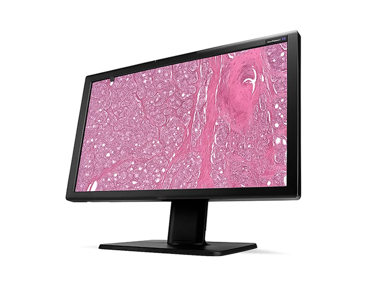
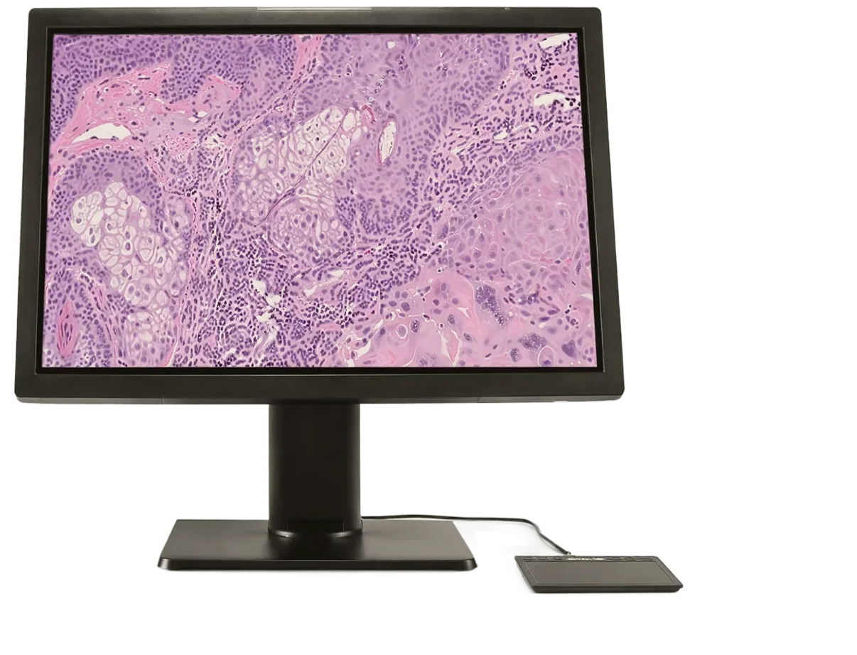
Key elements of the digital pathology environment
- Slide scanner — converts tissue images on glass into high‑resolution digital files (up to 10 gigapixels).
- Viewing software — provides zooming, annotations, comparison, and image analysis.
- Display — the key visualization device that shows digital slides with accurate colors, high sharpness, and contrast.
- Storage — PACS systems or specialized archives for long‑term storage and collaboration.
Technologies that matter
Displays for digital pathology
The display is the central element of the digital pathologist’s workstation.
It must convey the slightest differences in tissue shades while maintaining accuracy during magnification, scrolling, and image comparison.
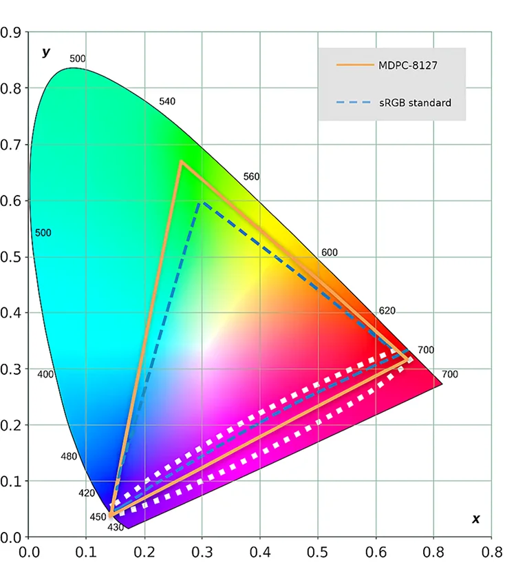
Unlike diagnostic or radiology screens, displays for digital pathology have a shifted color gamut (pathology mode) — warm pink‑beige tones that provide natural perception of stained histological specimens.
Accurate color calibrations to sRGB and GSDF, high pixel density, and uniform backlighting allow the pathologist to see color nuances of tissues — from weak cytoplasmic staining to saturated areas of the nucleus.
RapidFrame™
A technology that eliminates image blur during movement and zooming.
Fast frame refresh ensures smooth navigation through digital slides and high detail clarity even when working with large images.
Benefits:
- No ‘smearing’ when scrolling and zooming;
- Fast pixel adaptation when changing scale;
- Increased analysis efficiency and diagnostic speed.
Uniform Luminance Technology™
Uniform backlighting technology across the entire screen area.
It eliminates brightness and contrast variations, ensuring stable color perception across the entire image.
Especially important when evaluating large slides where color transitions must remain uniform.
Ambient light compensation
Built‑in sensors analyze the level of external illumination and automatically adjust image brightness and contrast.
This ensures visualization accuracy regardless of laboratory conditions.
SlideRightQA™
Artificial intelligence‑based software for quality control of digital slides.
It automatically detects defects — defocusing, missed areas, skew — and sends only correct images for analysis.
This reduces preparation time, increases accuracy, and reduces the workload on specialists.
QAWeb Enterprise
An intelligent display calibration and control system.
It automatically performs quality tests, checks brightness, contrast, and color reproduction, ensuring stable image quality throughout the display’s lifetime.
Certified displays for digital pathology
Barco MDPC-8127 — the first standalone display certified for digital pathology (IVDR and FDA)
It provides:
- 8 MP resolution,
- accurate color reproduction,
- built‑in sensors for automatic calibration,
- five‑year warranty,
- compatibility with most WSI and PACS solutions.
Also the Eonis 8MP display supports a special pathology mode with sRGB calibration optimized for the color characteristics of histological specimens.
Application
- 8 MP resolution,
- accurate color reproduction,
- built‑in sensors for automatic calibration,
- five‑year warranty,
- compatibility with most WSI and PACS solutions.
Why this matters
The growing volume of studies and the shift toward personalized medicine require new tools for data analysis.
Displays for digital pathology provide high image quality, accelerate diagnostic processes, and create a foundation for the integration of artificial intelligence technologies.
Recommended products
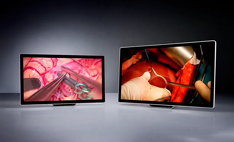
Surgical Displays
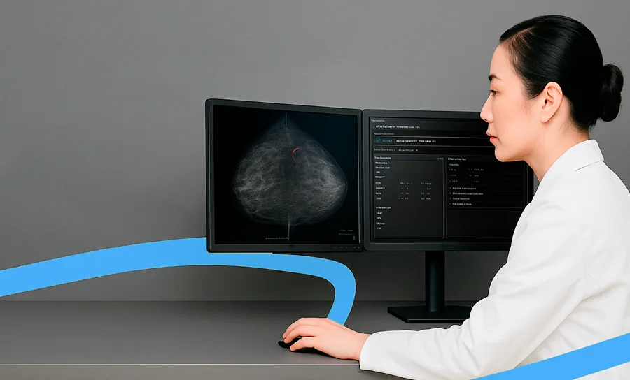
Medical computers
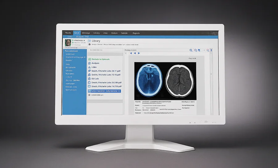
PACS/RIS solutions

Medical IT solutions
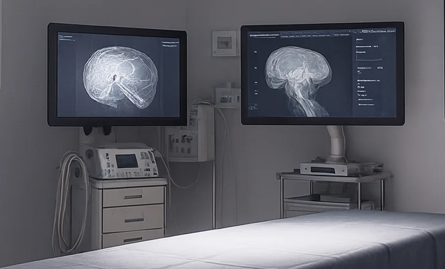
Digital Operating Room
Surgical Displays
Medical computers
PACS/RIS solutions
Medical IT solutions
Digital Operating Room
Surgical Displays
Medical computers
PACS/RIS solutions
Medical IT solutions
Digital Operating Room
Surgical Displays
Medical computers
PACS/RIS solutions
Medical IT solutions
Digital Operating Room
Technologies that make medicine more precise
The ITCO MED team selects digital pathology solutions tailored to the specific needs of laboratories and research centers, organizes demo testing, integration with WSI systems, and specialist training.